


When dealing with complex post-surgical orthopaedic and neurological cases, clinical observation alone is often not enough to fully capture the extent of dysfunction or recovery. Subtle compensations, neurological deficits, and
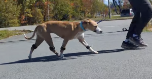
Functional assessment and rehabilitation monitoring using EKICO Tendiboots™ following surgical management of Traumatic Fragmented Medial Coronoid Process (Jump-Down Syndrome), a clinical case presented by Fur Ability Animal Rehabilitation in collaboration
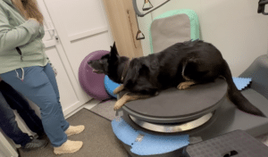
In this clinical case, Dr. Gudrun Werner presents the longitudinal management of Mila, a 10-year-old German Shepherd diagnosed with lumbosacral instability and spondylosis. Through repeated gait analysis with Tendiboots™


