Diagnosis of Hip Dysplasia and Sacrum Malformation in Sport Dogs.
How Can Tendiboots™ Canine Locomotion Data Be Useful in Diagnostic Research?
From Sensation to Diagnosis
February 2024
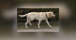
Example of a Use Case for the Tendiboots Canine Tool on a Female Dog Collected in February 2024.
Piv, a small female dog, trains and competes in agility competitions. At the time of our assessment, the owner did not recognize any locomotion issues in the dog, and Piv was actively participating in a training session.
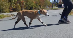
On the day of data collection, the analysis results from the Tendiboots™ Canine system highlighted several parameters outside the normal range and typical standard deviations observed in healthy dogs.
According to the data, the system indicated that the dog exhibited asymmetries that had not been reported by the owner.
On the day of analysis, the Tendiboots™ Canine system revealed an overload on the right forelimb and a reduced impact force on the right hindlimb.
The comparison of stride lengths and heights between the hind limbs also indicated an underutilization of the right hindlimb.

April 2024
In April, Piv’s owner consulted her veterinarian because the dog exhibited visible asymmetries in her locomotion.
An intermittent lameness, vaguely defined by her owner and the entire agility club to which she belonged, was noted.
Something was amiss in the small dog’s movement, though it was not clearly identified, prompting a consultation with her treating veterinarian, Dr. Valérie Foucon.
At that time, the veterinarian requested the data collected in February.
Subsequently, the dog clearly demonstrated an asymmetry in her locomotion, limping on the right hind limb.
Examinations conducted led to a diagnosis of right hind limb hip dysplasia along with sacral malformation.
A first sacral vertebra functioning like a lumbar vertebra.
The lumbar region of the vertebral column in dogs consists of seven vertebrae, while the sacrum is made up of three sacral vertebrae fused together to form a single bone, the sacrum. This fusion of the sacral vertebrae strengthens the structure and provides stability to the pelvis, which is essential for the movement of the hind limbs and for effectively transmitting forces during locomotion.
In the case of the dog Piv, the first sacral vertebra is not fused with the second sacral vertebra. This malformation of the sacrum effectively gives her the equivalent of an “eighth lumbar vertebra” instead of seven, meaning her sacrum consists of only two vertebrae instead of three.

This anomaly in the spine leads to instability in the pelvic region, which, along with hip dysplasia, explains the lameness observed in her locomotion. The pain may be further exacerbated by the fact that the sciatic nerve passes through the spaces formed by the lumbosacral vertebrae.
Hip Dysplasia in Dogs

Hip dysplasia is characterized by poor formation of the hip joint, resulting in an improper fit between the head of the femur and the acetabulum of the animal.
The primary consequences of this condition include chronic pain due to inflammation of the joint.
These abnormal frictional forces can lead to decreased mobility and muscle atrophy over time. Premature osteoarthritis is also frequently observed.
Ultimately, this issue affects the dog's quality of life, often limiting its ability to move.