One of the most commonly encountered joint diseases in dogs is osteoarthritis – a progressive, irreversible degeneration of the joints that leads to pain, lameness, stiffness, and loss of mobility.
While sporting dogs are more prone to this condition, all dogs can be affected, regardless of whether they engage in intense or light physical activity. This disease is also known as osteoarthrosis.
How to Detect Osteoarthritis?
First of all, it is important to understand that the degeneration that leads to osteoarthritis affects the entire joint structure, including the cartilage, synovial membrane, joint capsule, and subchondral bone.

Detecting this condition as a dog owner can involve noticing several different situations: the dog may show changes in behavior during physical activity (difficulty jumping, moving, etc.); at home (trouble climbing stairs or jumping onto the couch); and even during walks (limping, stiffness, etc.).
Changes in the animal’s behavior can also be associated with increased aggression or, conversely, apathy. Other clinical signs may also be present, such as a reduced appetite or visible signs of pain (whining, discomfort when being handled, or even pain upon touch).
In some cases, muscle atrophy may also be observed. One or more joints may be affected, and these symptoms can sometimes appear intermittently rather than being constant.
There are two main types of osteoarthritis: primary and secondary.
Primary osteoarthritis is often related to age, genetic predisposition, or natural joint wear over time.
Secondary osteoarthritis can result from mechanical stress, joint disease, osteochondritis dissecans (OCD), excess weight, or obesity.
A clinical example of how mechanical joint stress affects locomotion is illustrated in our case on the management of carpal hyperextension in dogs
It is important to note that secondary, mechanical osteoarthritis is the most common type in dogs. It typically develops as a consequence of significant mechanical stress and/or improper distribution of forces on the joint cartilage.
How to Diagnose Osteoarthritis and at What Stage?
Collecting the medical history and background information is an essential step in diagnosing osteoarthritis in dogs. This includes understanding in which situations clinical signs appear (e.g., during physical activity, rest, or specific movements), whether physical activity is affected, and if any joint injuries or trauma have occurred in the past. Focusing on these circumstances is a fundamental part of the diagnostic process.
These initial findings should be followed by a comprehensive clinical examination, including joint palpation and mobilization, to rule out other possible causes.
Finally, additional tests, particularly X-rays, should be performed to confirm the diagnosis. Osteoarthritis is typically classified into four stages, ranging from Stage 1, with minor joint involvement, to Stage 4, with severely reduced joint mobility.
Additionally, a standardized method exists to clinically grade osteoarthritis in dogs: the COAST (Canine Osteoarthritis Staging Tool). This tool allows both the veterinarian and the pet owner to assess the quality of life of the arthritic dog.
In a second step, the veterinarian also evaluates the severity and progression of osteoarthritis through clinical examination and additional tests, including radiographs. A score is then assigned to the dog, which not only confirms the diagnosis but also allows for ongoing monitoring and treatment adjustments as needed.

What Are the Challenges in Diagnosing Osteoarthritis?
All dogs can be affected by osteoarthritis.
Many factors come into play in the diagnosis (age, breed, physical activity, etc.), but each dog reacts differently to this condition.
The clinical presentation of osteoarthritis is non-specific, and other diagnostic hypotheses should sometimes be considered depending on the clinical signs presented by the animal, its medical history, and background information.
In its early stages, this condition may not be immediately symptomatic. One dog may start showing clinical signs early, while another may develop symptoms much later.
This diagnostic challenge is well illustrated in our clinical case exploring hidden carpal pain with the Tendiboots™ Canine, where subtle joint discomfort was only revealed through objective locomotion data.
Additionally, the variability of clinical signs must be taken into account. This is why joint X-rays are essential for a proper diagnosis. An analysis of synovial fluid through arthrocentesis can also help detect inflammation and is useful for differential diagnosis.

Why Detect Osteoarthritis Early?
Overall, the quality of life of a dog with osteoarthritis can be more or less affected, depending on the severity of the condition. The goal of early diagnosis is to prevent the onset of pain and other clinical signs that impact the dog's well-being, as well as to slow the progression of the disease.
The irreversible nature of osteoarthritis is also a critical factor. The earlier treatment is initiated, the lower the risk of severe joint damage. Early intervention also allows for better assessment of the treatment's effectiveness, enabling adjustments as needed.
For sporting dogs, which are particularly susceptible to this condition due to the potential development of micro-cartilage lesions, the absence of early treatment can lead to irreversible locomotor impairment and significant pain.
Additionally, the sporting career of the dog can be compromised if appropriate management is not implemented in time.
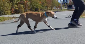
Early Detection of Osteoarthritis in a Sporting Dog with Tendiboots™ Canine
During a training session with a sporting dog, quantitative locomotion measurements were taken using the Tendiboots™ Canine system. Data was collected at the beginning of the session, throughout various exercises (jumping, attack, backing up), and at the end of the session. This evaluation, conducted on a Malinois involved in ring sport, lasted a total of 50 minutes.
The recorded data revealed a progressive asymmetry in the distribution of weight between the limbs as the exercises progressed.


Unexpected Results Despite a Normal Appearance
Although no visible abnormalities were observed at the start of the session, and no signs of lameness had been noted in the days leading up to it, the data collected by Tendiboots™ Canine revealed a progressively uneven distribution of weight, worsening as the training session continued.
Start of Session (Image 1)
At the beginning of the session, the weight distribution values were relatively balanced, ranging from 21.3 N/kg to 22.3 N/kg (areas highlighted in yellow).
End of Session (Image 2)
By the end of the session, significant discrepancies were observed, ranging from 16.5 N/kg for the left hind limb to 32.2 N/kg for the right forelimb.
When comparing the forelimbs to each other and the hind limbs to each other, it becomes clear that the values for the left side are consistently lower than those for the right side:
– 29.5 N/kg for the left forelimb versus 32.2 N/kg for the right forelimb
– 16.5 N/kg for the left hind limb versus 21.9 N/kg for the right hind limb
An Increasing Asymmetry Revealed by Dynamic Data
In the graph (Image 3), the values gradually drift away from the central horizontal line as the session progresses, revealing a growing asymmetry over time.
Diagnosis
Although the animal showed no visible signs of discomfort, X-rays taken after the session revealed early-stage osteoarthritis in the left hip, confirming the value of objective data provided by Tendiboots™ for the early detection of locomotor issues.
A similar diagnostic pathway is described in our clinical case on hip dysplasia and sacrum malformation in dogs, where gait data helped uncover underlying hip pathology.
In Conclusion
The management of osteoarthritis should be multimodal and include a comprehensive action plan.
Each dog should be treated on a case-by-case basis, with an individualized approach. Recently, monoclonal antibodies have entered the market as a promising treatment option, offering pain relief for dogs suffering from this joint condition.
In addition to pain management, the dog's physical activity must be appropriately adjusted. One common mistake owners make is to completely eliminate physical activity, when in fact it should be maintained, but properly dosed.
Discontinuing all physical activity can lead to several complications, such as muscle atrophy or ankylosis (joint stiffness).
Beyond medication (NSAIDs, corticosteroids, etc.), other disciplines can play a critical role in a multimodal treatment plan, including massage therapy, physiotherapy, dietary adjustments, leash walks, and changes to the dog's living environment.
In some cases, surgical intervention may also be necessary.
FAQ
In its early stages, osteoarthritis can be asymptomatic and escape visual examination, especially in a sporting dog. Tendiboots™ Canine dynamic analysis reveals a load-bearing asymmetry invisible to the eye. For example, in a ring-sport Malinois with no apparent lameness, recordings showed an initially balanced weight distribution (21.3 to 22.3 N/kg) that drifted as exertion progressed. X-rays taken afterwards confirmed early-stage osteoarthritis in the left hip, validated by the objective data.
A load asymmetry may only appear after exertion, when fatigue unmasks the painful area. In the Malinois case, values were balanced at the start of the session but showed a marked gap by the end: 16.5 N/kg on the left hindlimb versus 21.9 N/kg on the right, and 29.5 versus 32.2 N/kg on the forelimbs. Measuring locomotion across a working session (here 50 minutes) objectifies a progressive drift that a static at-rest examination would miss.
Per-limb peak vertical ground reaction force (GRF) is the central marker: comparing left/right forelimbs and left/right hindlimbs reveals one side consistently less loaded. On the progression graph, values gradually drift away from the central line as the session advances, reflecting growing asymmetry. These reproducible quantitative data turn a clinical impression into a comparable measurement, usable for differential diagnosis and follow-up.
Osteoarthritis is a progressive, irreversible degeneration: the earlier management begins, the lower the risk of severe joint damage. In the sporting dog, exposed to cartilage micro-lesions, the absence of early treatment can lead to irreversible locomotor impairment and compromise the dog’s career. Detecting a subclinical asymmetry under exertion, before overt signs appear, allows earlier intervention and subsequent objective assessment of treatment efficacy.
Primary osteoarthritis is related to age, genetic predisposition or natural joint wear. Secondary osteoarthritis, the most common form in dogs, results from mechanical stress, joint disease, osteochondritis dissecans (OCD), excess weight or obesity, through improper force distribution on the cartilage. Identifying a load asymmetry on gait analysis points to a mechanical component and helps target the cause to correct