Specialized in the follow-up of the sport horse - in particular dressage -, Pierre Martinuzzi has agreed to tell us how he takes care of horses suffering from common foot problems, benign or not, and sometimes requiring the assistance of an equine veterinarian.
Negative palmar angle
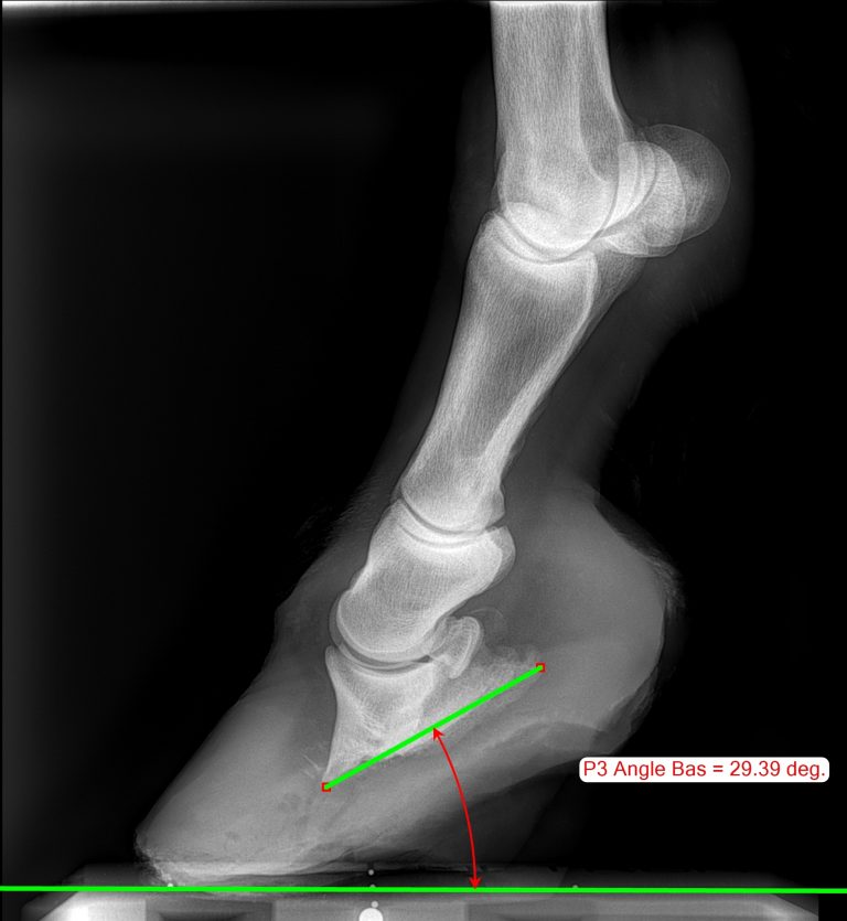
Often associated with flat hoof (in Thoroughbreds, for example), the retroversion of P3 also refers to the negative palmar/plantar angle. The distal phalanx (P3), which is located in the hoof capsule, respects a slight inclination in a so-called “normal” position.
This value is established by the examinations carried out by the referring veterinarian in charge of monitoring the horse and may vary from one horse to another. For example, it can depend on many factors: the horse's lifestyle, activity, history, conformation, etc.
On an X-ray, an angle considered normal for P3 (angle formed between the bottom of P3 and the sole) is generally between 4 to 7 degrees. Seen from the outside, a toe angle considered normal is generally between 47 and 53 degrees. As mentioned above, these indicative values must be adapted and contextualized for each horse by qualified professionals.
This last phalanx can nevertheless also have an abnormal position innately, as in the case of a malformation such as clubfoot for example, or acquired in the case of pathologies such as laminitis. These abnormal positions have repercussions on the locomotion of the horse and can cause pain and lameness. In the case of retroversion of P3, veterinarians and farriers note on the X-ray that the angle becomes negative. The angle of the last phalanx can drop below the horizontal in some cases!
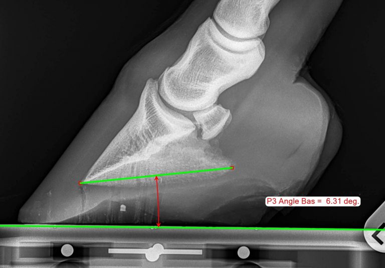

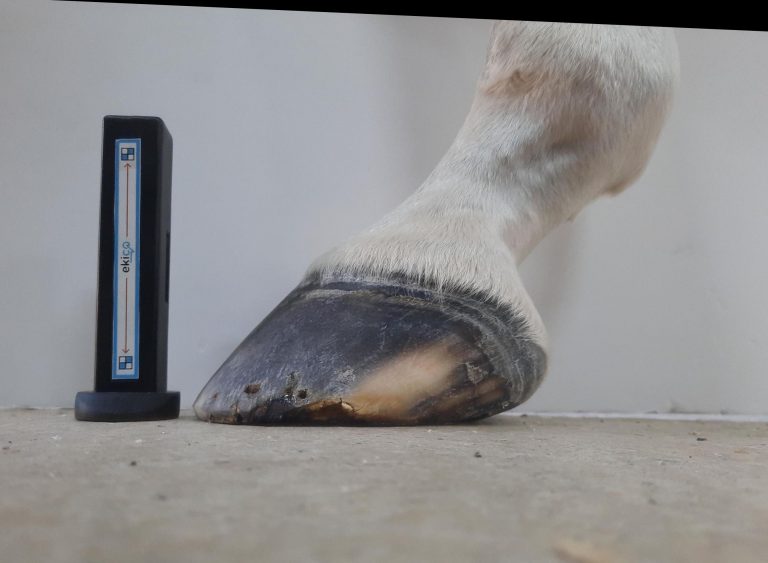
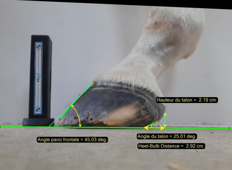
Conversely, the third phalanx can swing sharply and have a much too high angle, especially in cases of laminitis, for example.
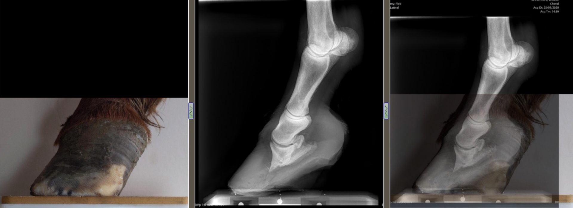
A word from the professional with Pierre Martinuzzi:
“In cases of retroversion of P3, collaboration between the veterinarian and the farrier becomes essential. X-rays of the feet and their analyzes make it possible to deal with the nearest millimeters to rectify the angle of P3.
In terms of shoeing, I generally achieve strong rolling on the outside edge of the shoe, toe inner to toe outer, for horses with a negative palmar angle. The recoil of the horseshoe allow not to slow down the start of the foot, reduces breakover and promotes heel regrowth by limiting their overload. Toe is therefore plunging in this type of configuration.
On some horses, the addition of a silicone support with leather plate is an additional help. As a farrier, the difficulty on this type of horse with a retroversion of the third phalanx is to estimate the amount of sole that can be removed during trimming.
The challenge is not to remove too much as this can easily cause discomfort for the horse. In such cases, working with objective measurements like Metron is a real help for professionals.”

Cracks
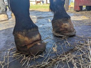
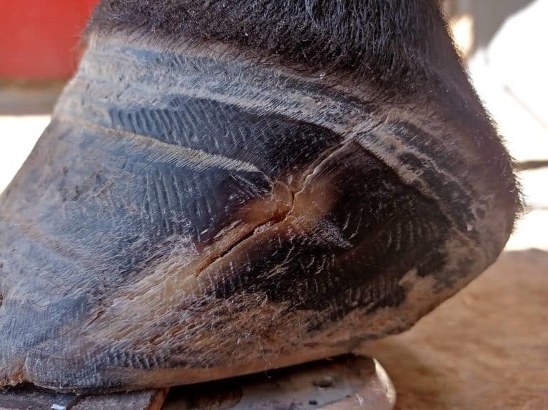
The crack is caused by a bad distribution of the loads on the hoof capsule. It is manifested by a bursting of the hoof on the surface but can also affect the soft tissues. In cases where the hoof cracks up to the coronary band, the horse may experience severe pain because, in motion, the soft tissues can get stuck between the hard tissues and cause painful pinches with each stride.
Hooves can be affected by different types of cracks. One distinguishes in particular the crack of meadow horses from the crack of sport horses regularly monitored. The crack of meadow horses generally starts from the sole and is often due to the negligence of owners who do not have their horses trimmed regularly. The excess hoof growth simply bursts over time! Luckily, the crack rarely goes up to the coronary band because the hoof does not break, due to its hardness. This type of crack therefore generally has little impact on the horse.


The crack of sport horses, as for it, starts mainly from the middle of the hoof and affects the horses which are regularly monitored. This can be caused by poor shoeing, but its root cause results from a poor distribution of the loads on the hoof, for example in the case of compensations.
For various factors, a horse can overload one hoof more than the other. In this case, a crack can appear at the location of the most fragile part of the overloaded hoof. This area generally extends from the end of the toe (outer or inner) to the heels.

A word from the professional with Pierre Martinuzzi:
“The challenge for hooves with a crack is to reduce the excess load on the concerned area. The follow-up of the evolution of the hoof capsule is in this case mandatory. In terms of care, the rider / owner unfortunately has a relatively limited field of action apart from daily monitoring of the horse and warning his farrier if the crack comes to go up towards the coronary band.
To solve the problem, it is mainly up to the farrier to work to improve the distribution of loads over the entire hoof. Hoof parameters must be monitored with the utmost precision to ensure that loads and heel height do not increase, especially for horses with a so-called club foot morphology. There are a number of strategies that the professional can use to manage the cracks, prevent them from getting worse and bring comfort to the horse.”
For example, by having recourse to the removal of support (see pictures above), the area affected by the crack is then found in a vacuum to avoid any load. “Beveling in whistle in the lower region of the hoof will also remove some of the support and reduce the pressure exerted on the area affected by the crack. An effective method is also to staple the hoof when possible. The staples limit the separation of the hoof wall, prevent the opening of the crack and slow down its propagation towards the coronary band. The poor distribution of loads unbalances the hoof as a whole and has strong negative repercussions on the well-being and performance of the equine.”


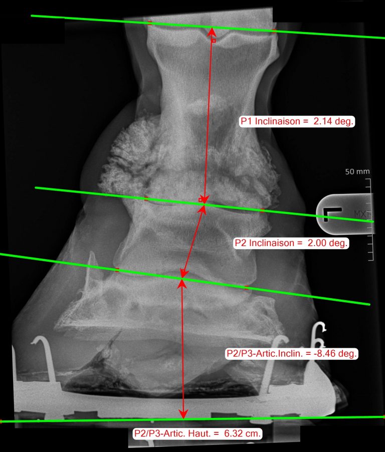
As a farrier, the difficulty is to reorganize the distribution of the loads on the foot without causing too much transfer to another part of the hoof. The professional therefore seeks to keep the hoof as flat as possible without P3 sagging on one side or the other. Hoof X-rays are extremely helpful in this case to ensure that the third phalanx does not collapse either medially or laterally.
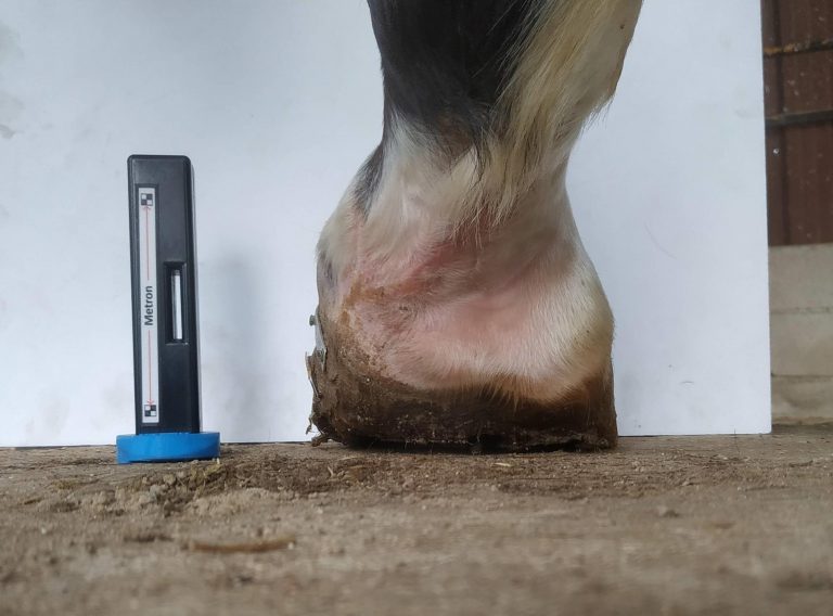
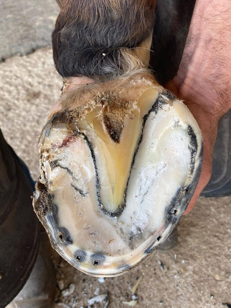
Metron’s measurements allow great accuracy to monitor the orientation of P3 in the hoof capsule, whether forward, backward, or sideways. This level of accuracy makes it possible to quickly realize the first millimeters of sagging and to react as quickly as possible before the situation becomes too serious for the horse.


This article was written in collaboration with Pierre Martinuzzi, farrier specialized in the monitoring of sport horses in collaboration with veterinarians in Paris area, France.
Find all his news on his Facebook page


