François Halbout, farrier located in Normandy (27) is specialized in the management of serious orthopedic pathologies such as: laminitis, navicular syndrome, clubfoot, hyperlaxity in foals, etc.
He gives us his time to share his experience with the management of severe laminitis cases.
Overview of a complex pathology that is still poorly understood.
Laminitis at a glance
Laminitis is a serious generalized disease, which affects the whole body and is expressed at the level of the horse’s feet. It causes an abnormality of blood circulation in the feet producing necrosis of the laminae.
In the most advanced cases, the coffin bone, pulled by the flexor tendon of the finger, can end up puncturing the sole.
The causes can be:
- Food: nitrogen / glucose imbalance in the blood, absorption of a large amount of cold water
- Mechanical / traumatic: overwork, chronic overload
- Medicinal: intoxication
- Infectious (parturition)
- Cushing syndrome and disease
Clinical signs and treatments
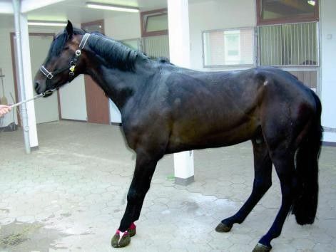
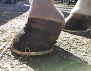
The horse is at a standstill, stands camped and puts all its weight on its heels. He moves little and the lameness is worse on hard ground. The pain is severe and may cause the horse to maintain a lying position.
As a first step, if the laminitis is detected very early, I recommend dipping his feet in ice to limit the symptoms and then reduce the pain with non-steroidal anti-inflammatory drugs. The horse must be relieved as quickly as possible. Personally I use 8 centimeters thick extruded polystyrene plates which have the advantage of being able to be put in place very quickly and without constraint for the horse, this will allow to provide a minimum of comfort and to limit the rocking and / or the collapse of the coffin bone.
Secondly, only after the end of the acute phase of laminitis (around 10 days), under no circumstances should the horse be shoeed in a conventional manner, that is to say, forcing him to stand on three limbs. This creates too much overload. It is best to shoe it lying down or suspended in a harness.
Specific trimming then becomes essential. The objective is to :
- lower the heels as much as possible to try to re-horizontalize the coffin bone.
- strongly grate the forceps parietal to align it with P3 and evacuate the necrotic podophyllum, thus limiting the formation of abscesses.
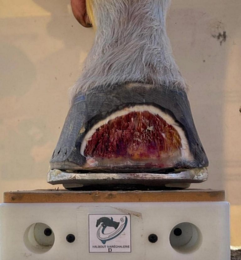
All of this well-controlled trimming coupled with a three-dimensional aluminum Banana style iron will replace the center of pressure.
It will thus be possible to re-establish vascularization in the foot and avoid necrosis of the apex of the distal phalanx. Obviously, they will be glued with resin and not nailed to avoid shocks due to broaching. To limit the tilting of the third phalanx and the distribution of loads, a very firm silicone (Luwex laminitis) or resin (Vettec) will be applied under the foot. The horse, for its part, must be kept in the box on thick bedding.
Another alternative is the installation of a screwed wooden iron which also effectively relieves the horse. I prefer it for very painful horses with very damaged feet and with uncontrolled abscess risks. This type of remedy makes it possible to maintain the three-dimensional effect. In addition, the absence of glue prevents the creation of heat which could cause any abscesses to flare up.

Pitfalls and difficulties associated with laminitis
1. The use of radio images only
From my experience, the single use of the radio gives partial information on the condition of the horse. In some cases this can be misleading.
To be more accurate, the radio provides important information about the current condition of the foot, some information on the past condition, but very little information on the incoming condition.
It is therefore common for a horse which was doing very well (“radiologically”) when the X-ray images were taken with little or no tilting or collapse of P3, declares a critical situation 3 weeks later.
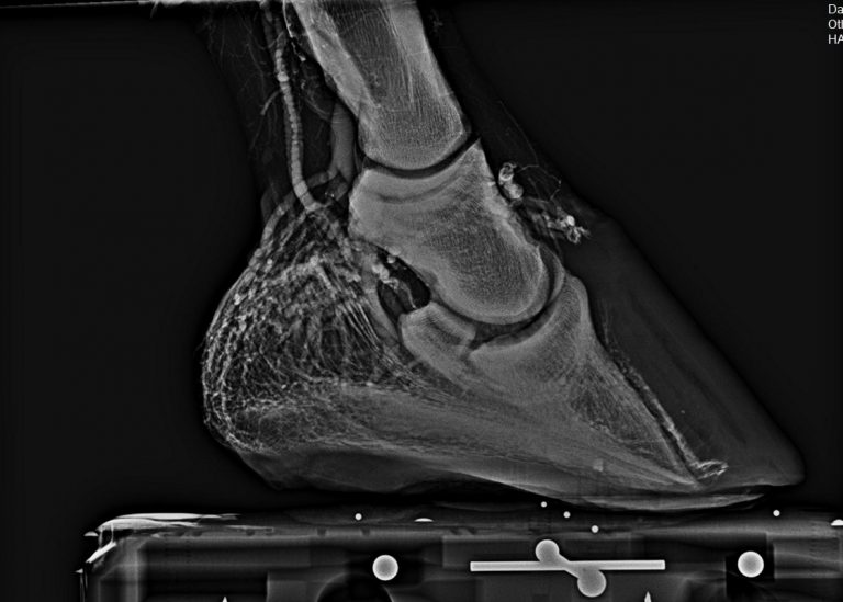
The phlebogram, for its part, makes it possible to quickly realize the vascularization defects of the foot. and realize that the problem will be more serious than what is currently seen on the radio. This examination is often closer to the observed clinic.
2. Rotation vs Descent
Some professionals focus a lot on the phalanx tilt and may forget to check for a descent which is ultimately much more devastating.
For this type of verification, the Metron software is particularly useful to me !
3. Shoe too quickly
From my experience, it is imperative to wait until the end of the acute phase before referral.
A laminitis attack causes significant inflammation in the foot. Any handling that is too rapid risks increasing this inflammation (trimming, change of supports, shocks, loading of the opposite foot, etc.) and consequently, making the animal more and more painful.
4. Tendon retraction
Tendon retraction is one of the complications that can set in as a result of poorly treated laminitis.
It is very common with Shetland. Less with horses which are generally more followed. They are also less resistant to pain and more rarely arrive at this stage of complication.
As a farrier, the reflex is to leave the heel behind because horses with laminitis find more comfort there.
This is a mistake becauseone of the possible consequences is to enter another vicious circle, and see a tendon retraction set in. The outcome can be just as fatal for the animal (see article “The essential role of the farrier by Bart Lambert“).
5. The relationship with the owners
A main difficulty encountered with owners of exhausted horses is the psychological aspect.
I always warn owners that the hardest part will be going through the next few months with their horses in pain, lying down.
It takes mental stamina, they don’t always realize it and want to get over the situation as quickly as possible.
Laminitis is a pathology from which it is difficult to get out, and it takes time.
In my opinion it takes a year for the horse to fully recover of this type of disease.
Of course, the horse will be better during this given year. But going too fast at the first signs of improvement increases the risk that the horse will not fully recover or that chronic laminitis will develop.
The financial aspect also often catches up with the emotional aspect, because specific treatments and care can be heavy to bear financially to keep the most serious cases in comfort.
Useful tools and technologies in laminitis management
Radiography coupled with venography are in my opinion essential to manage cases of laminitis.
Therefore, the veterinarian / farrier duo is essential.
The past emotion linked to the diagnosis, the owners do not always understand it, and hope to be able, over time, to save the visits / pictures taken by the veterinarian.
This is a serious mistake, because as a farrier, it pushes us to work partly blind. And on this type of pathology, the consequences can be dramatic.
The veterinarian / farrier duo is essential to manage laminitis cases
François Halbout
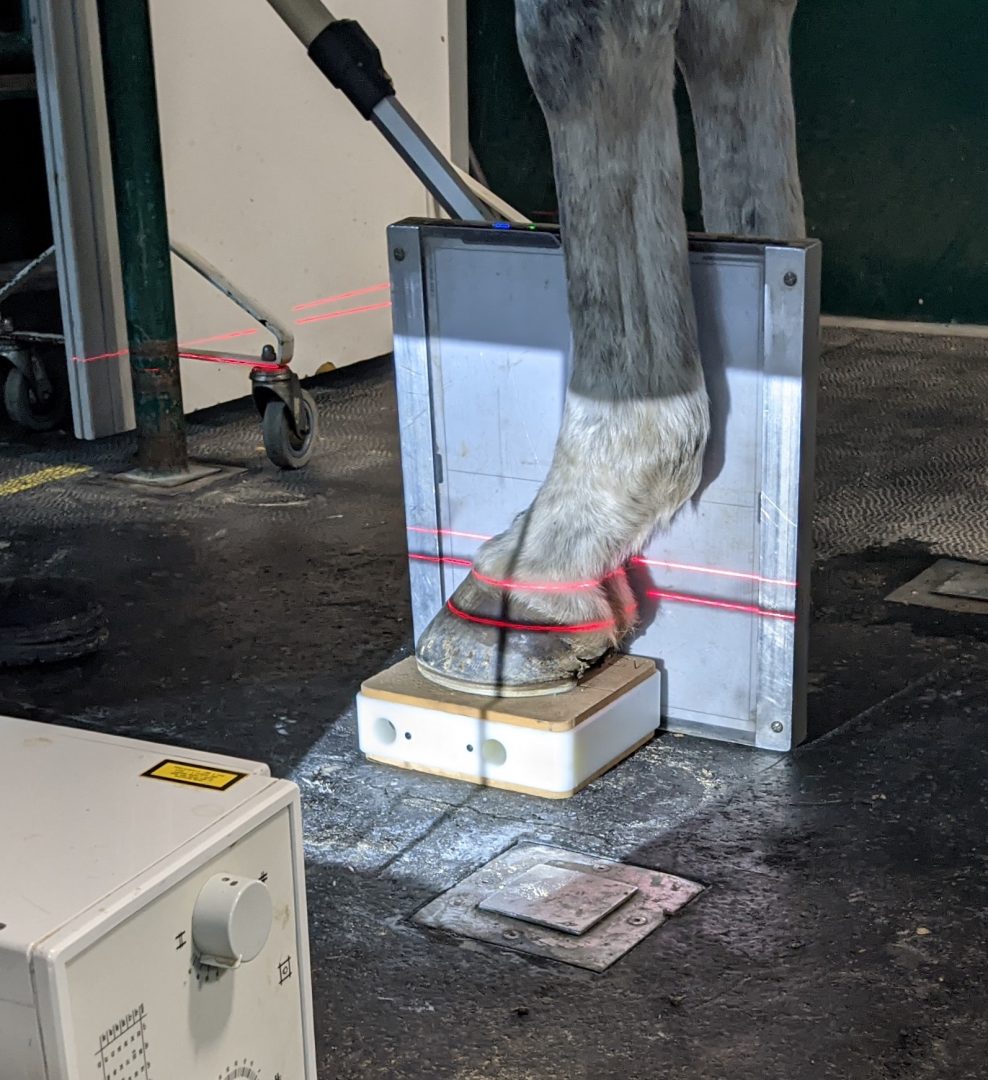
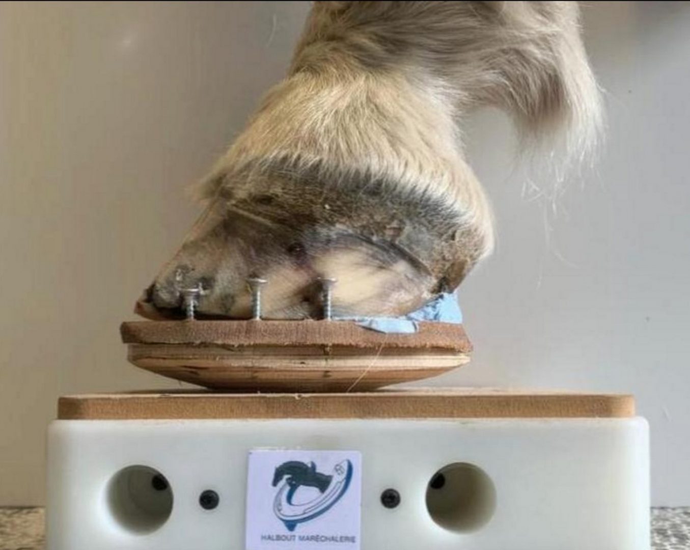
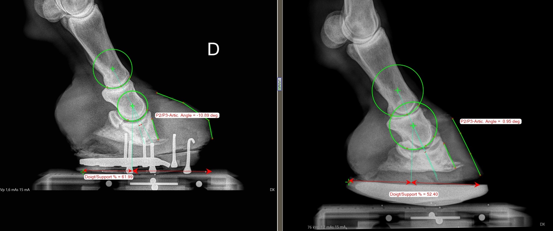
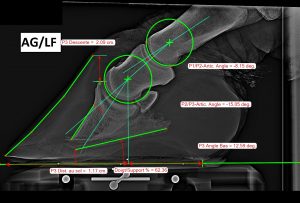
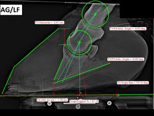
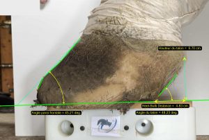
For my part, I also integrated the measurement into my activity.
I stay very close to the vets with whom I collaborate. From their photos I can follow things that are important to me as a farrier.
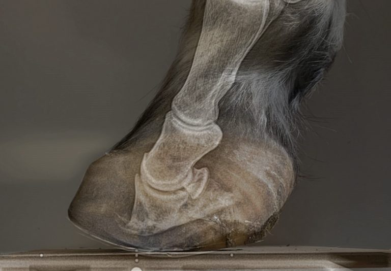
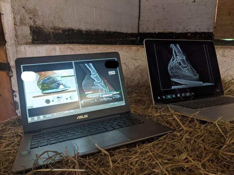
In some cases, I couple the veterinary x-rays taken on my blocks with my photographic images.
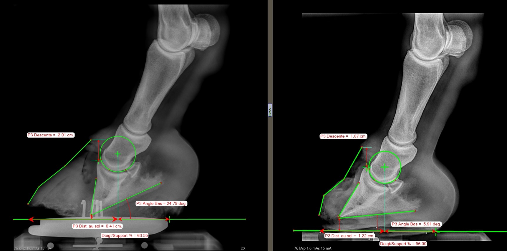
This allows me to gain in precision and autonomy, I have numerical proof that the horse will be better clinically. With the measurement, the improvements are quantified, more precisely than with the naked eye, it is indisputable, the measurement protocol remains the same from one shot to another, from one horse to the next to be as accurate as possible.
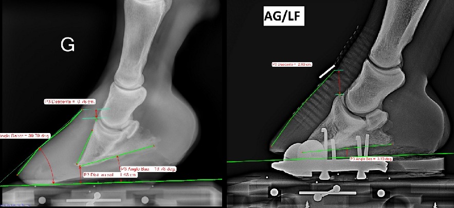
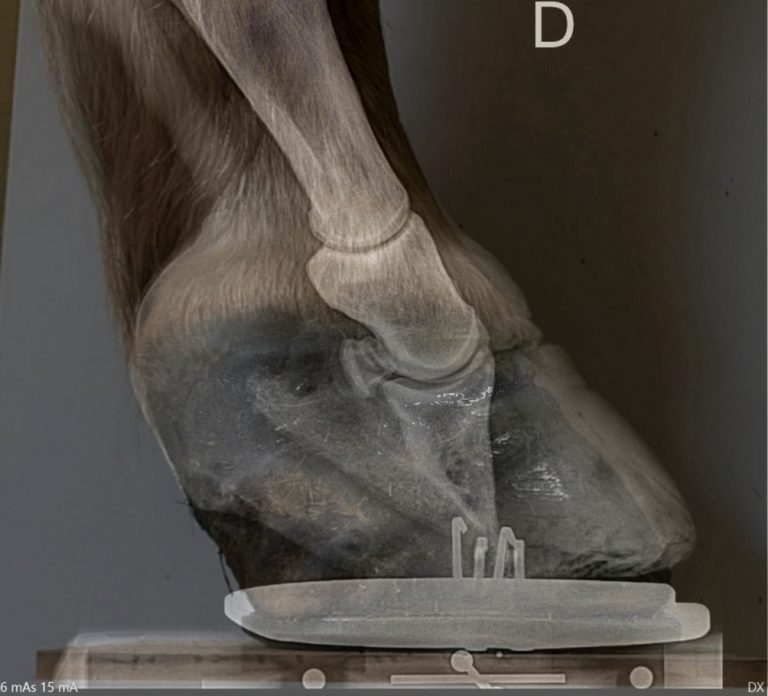
I use in particular the measurement of the phalangeal angles as well as the distribution of the distances on either side of the center of the condyle of P2.
With the measurement, the improvements are quantified, more precisely than with the naked eye, it is indisputable, the measurement protocol remains the same from one shot to another, from one horse to the next to be as accurate as possible.
François Halbout

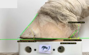
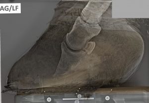
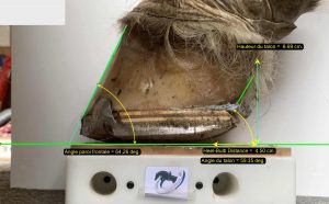
For the shoeing part, seeing the evolution at the exit of the shoe is a good thing to illustrate to my clients how the horse evolves as well as the result of the work done.
The photo reminds the owners how the horse was when they brought it to me, as they don’t always remember the horse was in this condition when I first picked it up!


